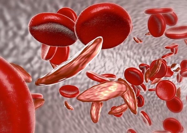
Sickle cell disease is a genetic condition that results in mutated forms of hemoglobin, the protein that carries oxygen to tissues and organs via the bloodstream. Mainly affecting those of Mediterranean and African ancestry, the disease can cause significant morbidity and mortality but the frequency and degree of the condition do tend to vary amongst individuals.
Unfortunately, there is no cure for sickle cell disease since it is a genetic disorder but there are therapies available to help manage patients who do develop symptoms.
Signs and Symptoms
Sickle cell disease usually presents early in childhood and there are numerous signs and symptoms that can develop due to the condition. These may include:
- Anemia – where patients may develop issues such as shortness of breath, heart palpitations, pale skin, dizziness, and even confusion as a result of the inability of the body to carry adequate amounts of oxygen to essential organs.
- Acute and chronic pain – sickle cells disease is associated with a vaso-occlusive crisis (where the blood vessels constrict) resulting in pain. These pain crises are the most distinguishing clinical feature of sickle cell disease.
- Aplastic crisis – this is a serious complication as a result of being infected with parvovirus B19.
- Growth and developmental delays such as delayed sexual maturation.
- Bone pain – as a result of obstruction of blood flow through the bone marrow of long bones.
- Involvement of the cardiac system – the condition may result in abnormal swelling of both the ventricles and the left atrium of the heart.
- Hemolysis – the abnormal blood cells may rupture resulting in the release of substances such as bile into the bloodstream and this may cause jaundice (yellow discoloration of the eyes and/or skin), as well as causing the urine to appear darker.
- Gastrointestinal involvement – children may develop gallstones due to the increased levels of freely circulating bile and the liver may also be affected.
Involvement of the Eyes
The eye-related issues that may develop due to sickle cell disease are vast and may involve multiple areas of the organs and these will be discussed further.1 As mentioned, sickle cell disease is a condition that results in constriction and obstruction of the blood vessels and this may occur in those supplying nutrients and oxygen to the irises, conjunctiva, choroids, and retinas.
Other conditions may also cause the same ocular changes that occur with sickle cell disease. Therefore, it is important to rule out disorders such as retinal vein occlusion, diabetic retinopathy, Eales disease, uveitis, polycythemia vera, and familial exudative vitreoretinopathy.
Anterior segment abnormalities
- Hyphema–where blood collects in the anterior chamber (space between the iris and cornea).
- Cataracts which occurs when the lenses of the eyes become dull and cloudy.
- Infarct and atrophy of the iris.
- Phthisis bulbi which is a shrunken and non-functional eye.
Posterior segment abnormalities
- Optic disk changes –occlusions of the intravascular vessels on the surface of the optic disk seem like dark red spots and don’t cause any clinical issues to the affected patient.
- Chronic macular changes due to microaneurysms developing in the blood supply to this tissue.
- Posterior retinal occlusions may involve the central or branching arteries and rarely the retinal veins. Macular arteriolar or peripapillary occlusions are also rare.
- Proliferative sickle retinopathy – this is the most severe ocular manifestation of sickle cell disease. The condition is associated with peripheral changes of the retina, especially in patients diagnosed with the hemoglobin SC variant of sickle cell disease. Those with hemoglobin AC, hemoglobin AS, homozygous hemoglobin SS, and hemoglobin D-thalassemia disease are also prone to develop this complication. Proliferative sickle retinopathy is a progressive disease and the ultimate objective is to treat the vascular tissue before a vitreous hemorrhage occurs which will compromise the patient’s vision. The five stages of peripheral sickle retinopathy are peripheral arteriolar occlusions, arteriolar-venular anastomosis, neovascular proliferation, vitreous hemorrhage, and then detachment of the retina.
- Non-proliferative retinopathy – also has a progressive nature to it and involves certain processes. Arteriovenous shunting occurs from the peripheral retina which may lead to venous tortuosity which results in bleeding inside the retina which causes marks to appear which are referred to as salmon-patch hemorrhages. When the intraretinal hemorrhage disappears, it forms a schisis cavity. A black sunburst lesion develops around the chorioretinal scars located around the equatorial fundus but these don’t cause any visual issues.
Reference
- Shukla P, Verma H, Patel S, Patra PK, Bhaskar LVKS. Ocular manifestations of sickle cell disease and genetic susceptibility for refractive errors. Taiwan Journal of Ophthalmology. 2017;7(2):89-93. doi:10.4103/tjo.tjo_3_17.


Be First to Comment