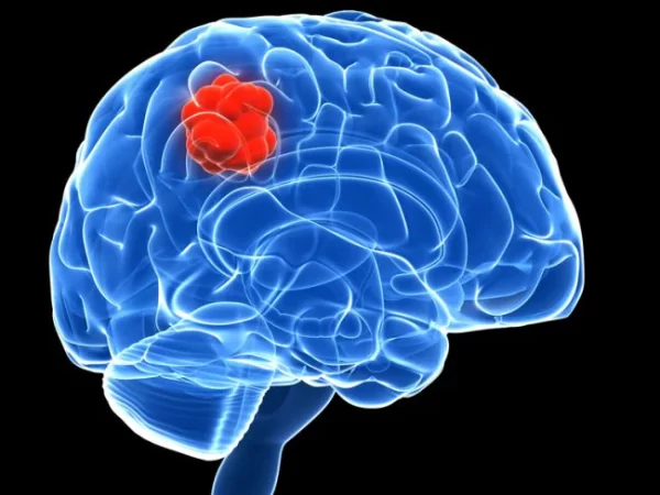
Gliomas represent a heterogeneous group of primary brain tumors that arise from the glial cells, which support and nourish the neurons within the central nervous system (CNS). Gliomas account for a significant portion of all brain tumors and can manifest across a broad spectrum of malignancies, ranging from benign to highly aggressive and malignant forms. The prognosis of gliomas gets deeply intertwined with the intricate molecular, genetic, histological, and clinical characteristics that define these tumors.
Classification and Molecular Markers
Gliomas are classified based on their cell of origin and molecular characteristics. The World Health Organization (WHO) classification system, revised in 2016, stratifies gliomas into various categories based on histological and molecular features. This classification is instrumental in guiding treatment decisions and estimating prognosis.
Diffuse Gliomas (WHO Grade II-IV)
Diffuse Astrocytic and Oligodendroglial Tumors (IDH-Mutant)
These tumors harbor mutations in the isocitrate dehydrogenase (IDH) genes, primarily IDH1 and IDH2. They are further categorized into oligodendro and astrocytomas, with IDH-mutant and 1p/19q co-deleted tumors indicating better prognosis due to increased sensitivity to therapies.
Diffuse Astrocytic and Oligodendroglial Tumors (IDH-Wildtype)
This subgroup includes glioblastomas (GBM), the most aggressive form of glioma. IDH-wildtype GBMs have a poor prognosis due to their resistance to conventional therapies and highly infiltrative nature.
Other Gliomas
Pilocytic Astrocytoma (WHO Grade I)
Generally, benign tumors most commonly occur in children and young adults. Surgical resection is often curative.
Ependymomas (WHO Grade II-III)
Arise from ependymal cells lining the ventricles and spinal cord. Prognosis varies widely based on location, grade, and extent of resection.
Histopathology and Grading System
Histopathological analysis plays a pivotal role in glioma prognosis. The WHO grading system categorizes gliomas into four grades (I-IV) based on histological characteristics, mitotic activity, necrosis, microvascular proliferation, and nuclear atypia. Higher-grade tumors are associated with worse outcomes. Glioblastoma (GBM), a WHO Grade IV glioma, is characterized by aggressive invasion, angiogenesis, and resistance to treatment.
Molecular Biomarkers:
IDH Mutations
IDH mutations are crucial for classification and serve as prognostic solid indicators. Patients with IDH-mutant gliomas generally have longer survival rates than those with IDH-wildtype tumors.
1p/19q Co-deletion
This genetic alteration is particularly relevant in oligodendrogliomas and signifies a better prognosis.
MGMT Promoter Methylation
Methylation of the O^6-methylguanine-DNA methyltransferase (MGMT) promoter predicts a better response to temozolomide chemotherapy in GBM patients.
TERT Promoter Mutations
Mutations in the TERT promoter are associated with increased telomerase activity and are linked to poor prognosis, particularly in high-grade gliomas.
Prognostic Factors and Clinical Presentation
Clinical factors impact glioma prognosis, including age, performance status, extent of surgical resection, and neurological deficits at diagnosis. Younger patients often have better outcomes due to increased tolerance for aggressive treatments. Additionally, the tumor’s location within the brain can influence prognosis, as tumors in functionally critical areas may be less amenable to complete resection.
Treatment Modalities and Their Impact on Prognosis
Surgical Resection
Maximal safe surgical resection is a cornerstone of glioma management. It alleviates mass effects, relieves symptoms, but also aids in obtaining tissue for accurate diagnosis and molecular profiling. The extent of resection, particularly for high-grade gliomas, significantly impacts survival. However, due to the infiltrative nature of gliomas, complete resection may not always be feasible.
Radiotherapy
Radiotherapy, primarily in the form of external beam radiation, is a standard treatment for gliomas. It targets residual tumor cells post-surgery and controls tumor growth. Fractionated radiotherapy regimes have demonstrated survival benefits, especially in high-grade gliomas.
Chemotherapy
Chemotherapy, often administered alongside radiotherapy, includes agents like temozolomide, which alkylates DNA and disrupts cell division. The MGMT promoter methylation status predicts response to temozolomide. Combination therapies, targeted agents, and immunotherapies are explored to improve outcomes.
Emerging Therapies
Targeted Therapies
Molecular profiling has enabled the development of targeted therapies against specific mutations, such as IDH and BRAF inhibitors, for particular subtypes of gliomas.
Immunotherapies
Immune checkpoint inhibitors and personalized cancer vaccines hold promise for glioma treatment by harnessing the body’s immune system to target tumor cells.
Gene Therapies
Innovative approaches, like chimeric antigen receptor (CAR) T-cell therapy, aim to genetically modify a patient’s immune cells to recognize and eliminate glioma cells.
Prognosis Stratification
The prognosis of gliomas is highly variable and depends on the intricate interplay of multiple factors, including molecular markers, histological characteristics, patient age, performance status, and treatment response. Predictive models and scoring systems are developed to predict outcomes more accurately.
The Recursive Partitioning Analysis (RPA)
Developed for glioblastoma patients, the RPA stratifies patients into three risk groups based on age, Karnofsky Performance Status (KPS), and extent of surgical resection. This system provides a baseline for assessing prognosis.
The Molecular-Modified Glioma Prognostic Index (mGPI)
Expanding on the GPI, the mGPI integrates additional molecular markers (TERT promoter mutation, MGMT promoter methylation) to stratify glioma patients further, enhancing predictive accuracy.
Where to Find the Best Prognosis
Medical Professionals
Medical experts, particularly oncologists, and neurologists specializing in brain tumours, are primary sources of glioma prognosis information. These specialists consider numerous factors to provide a personalized prognosis based on the type of glioma, location, grade, size, patient age, overall health, and response to treatment.
Diagnostic Imaging and Tests
Advanced diagnostic tools play a critical role in assessing glioma prognosis. Magnetic Resonance Imaging (MRI), Computed Tomography (CT) scans, and Positron Emission Tomography (PET) scans provide detailed information about the tumor’s location, size, and potential spread. Biomarker analysis and genetic testing can offer insights into the tumor’s molecular characteristics, guiding treatment decisions and prognostic predictions.
Cancer Centers and Research Institutions
Prominent cancer centers and research institutions specializing in brain tumors often offer comprehensive information about glioma prognosis. Institutions like the Mayo Clinic, MD Anderson Cancer Center, and Dana-Farber Cancer Institute provide detailed resources, research updates, and access to experts who can offer insights into the latest glioma diagnosis and treatment advancements.
Conclusion
Gliomas remain a formidable challenge in oncology due to their complexity, heterogeneity, and invasive nature. The prognosis of gliomas hinges on a multifaceted interplay of molecular alterations, histopathological features, clinical characteristics, and treatment responses. Advances in molecular profiling have ushered in a new era of personalized medicine, enabling more accurate prognosis estimation and targeted therapeutic approaches. However, the quest for improved outcomes continues, spurring research into novel treatment modalities, precision medicine, and innovative techniques that may one day unravel the intricate web of glioma prognosis and transform the landscape of glioma management.

