

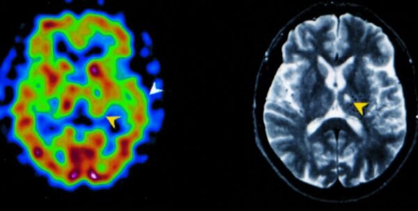
The Science Behind SPECT Imaging: Unraveling the Mysteries of Brain Function
What is SPECT Imaging and How Does it Work?
Single Photon Emission Computed Tomography (SPECT) is a powerful imaging technique that provides clinicians with detailed insights into brain physiology. Unlike traditional imaging methods, such as CT or MRI that primarily provide structural images, SPECT focuses on the functional aspects of the brain. It captures information about blood flow, metabolism, and overall brain activity. The technology uses radiotracers—a type of radioactive substance—that emit gamma rays as they decay. These tracers are usually injected into a vein and they circulate throughout the brain, binding to specific areas based on their metabolic activity. As the gamma rays are detected by the SPECT camera, they are converted into images that represent brain activity in real time. The process begins with the patient being prepared for the scan, which may include fasting or avoiding certain medications. Once the radiotracer is administered, there is typically a waiting period that allows the substance to distribute throughout the brain. The actual imaging takes a few minutes, during which the patient is asked to remain still. The resulting 3D images offer a visual representation of how different parts of the brain function, showcasing areas of normal activity and potential dysfunction. Ultimately, SPECT imaging provides clinicians with a unique perspective on neurological health that complements other imaging modalities.
The Role of Radiotracers in Mapping Brain Activity
Radiotracers are the cornerstone of SPECT imaging, essential for delineating physiological processes within the brain. A variety of radiotracers are utilized depending on the specific metabolic pathways or conditions being examined. For instance, Technetium-99m (Tc-99m) is one of the most commonly used radiotracers in brain imaging. It can target specific ligands that bind to receptors, allowing measurement of regional cerebral blood flow (rCBF) and metabolic activity. Different tracers can highlight specific neurotransmitter systems, providing valuable insights into neuropsychiatric conditions. For example, the tracers like [123I]FP-CIT are useful in assessing dopamine transporter availability, which can be crucial in diagnosing Parkinson’s disease. The ability to use these tailored radiotracers means that SPECT can adapt to various clinical needs, making it an invaluable tool in both research and clinical settings. Ultimately, the choice of radiotracers significantly influences the outcomes of the imaging study, allowing for increased specificity in diagnosing neural conditions.
Comparing SPECT Imaging with Other Neuroimaging Techniques
When considering neuroimaging techniques, it is essential to contextualize SPECT within the broader landscape of medical imaging. The primary methods include Functional Magnetic Resonance Imaging (fMRI) and Positron Emission Tomography (PET). While both fMRI and PET can provide functional information about the brain, they operate on different principles and have unique advantages and disadvantages. fMRI, for instance, relies on blood oxygenation level dependent (BOLD) contrast to assess brain activity. It is particularly effective for understanding functional connectivity across different brain regions, making it invaluable in cognitive neuroscience. However, fMRI has a lower temporal resolution than SPECT and may not effectively delineate rapid neural events. Conversely, PET imaging, similar to SPECT, utilizes radiotracers but typically employs positron-emitting isotopes, providing metabolic information that can be captured with high sensitivity. While PET imaging yields more quantifiable data, it is often more expensive and complex than SPECT. SPECT imaging stands out due to its cost-effectiveness, accessibility, and ease of use, making it especially advantageous in clinical practice. While it may offer lower spatial resolution than PET or fMRI, the rapid acquisition of data and opportunity for real-time monitoring of brain function allows SPECT to play a critical role in diagnosing and treating neurological disorders.
Applications of Brain SPECT Imaging: From Diagnosis to Treatment Planning
In the realm of neurology, brain SPECT imaging serves as a vital diagnostic tool, akin to a detective’s magnifying glass that uncovers hidden clues about a patient’s condition. Commonly, brain SPECT is employed to help identify conditions such as Alzheimer’s disease, Parkinson’s disease, and various forms of dementia. For example, patients with Alzheimer’s may exhibit distinctive patterns of reduced blood flow in regions like the temporal and parietal lobes, which are essential for memory and cognitive processes. In contrast, brain SPECT can assist in diagnosing frontotemporal dementia by revealing hypoperfusion in the frontal and anterior temporal areas.
Furthermore, this imaging modality plays a crucial role in differentiating between types of neurological disorders. It can help distinguish vascular dementia from neurodegenerative conditions by assessing blood flow patterns. Evaluating cerebral circulation allows clinicians to pinpoint areas affected by strokes or ischemic events, clarifying which regions are damaged versus those deteriorating as a result of disease. When traditional diagnostic methods prove inconclusive, brain SPECT offers a comprehensive and detailed perspective that supports more accurate and timely neurological diagnoses.
Assessing Brain Function in Psychiatric Conditions
SPECT imaging extends its utility beyond neurological disorders into the realm of psychiatry, where brain function can exhibit profound influences on mood, behavior, and cognition. One of the critical applications is in the assessment of conditions such as depression, schizophrenia, and anxiety disorders. The ability to visualize brain activity provides clinicians with insights that may not be observable through psychological evaluations alone. For example, research has shown that individuals with major depressive disorder frequently display decreased perfusion in the frontal lobes, which are associated with executive function, impulse control, and decision-making. Similarly, alterations in regional blood flow patterns can mark the presence and severity of psychotic symptoms in schizophrenia, allowing for tailored treatment planning. Moreover, SPECT imaging can uncover underlying neurobiological contributors to these psychiatric conditions, leading to a more nuanced understanding of patient behavior and responses to therapy. This rich data can guide physicians in choosing between various treatment options, monitoring changes in brain function over time, and adjusting therapeutic approaches to optimize patient outcomes.
Utilizing SPECT Imaging for Personalized Treatment Strategies
The era of personalized medicine continues to gain traction, and SPECT imaging plays a pivotal role in tailoring treatment to individual patient needs. Beyond diagnosing and assessing conditions, SPECT can assist in evaluating treatment efficacy by tracking changes in brain activity over time. For example, following administration of certain psychiatric medications, SPECT imaging can reveal alterations in cerebral blood flow patterns that signify changes in symptomatic relief or activation of neural pathways critical to mood regulation. Additionally, healthcare providers can utilize SPECT imaging data to develop patient-specific interventions. Armed with insights into how an individual’s brain reacts to stimuli or medications, clinicians can adopt approaches that resonate best with that patient’s unique brain physiology. For example, in patients diagnosed with conditions like Attention-Deficit/Hyperactivity Disorder (ADHD), SPECT imaging can reveal hypoperfusion in areas like the prefrontal cortex, allowing practitioners to create specialized and targeted cognitive-behavioral strategies and pharmacological options. Ultimately, SPECT imaging not only enhances diagnostic capabilities but actively contributes to the evolution of treatment protocols, ensuring comprehensive strategies adapted to the unique requirements of patients facing neuropsychiatric challenges.
Interpreting SPECT Images: The Art of Understanding Brain Maps
Reading Between the Lines: What SPECT Images Reveal
The interpretation of SPECT images involves a complex interplay between radiologists and referring physicians, an endeavor requiring a blend of medical expertise and analytical skills. SPECT results, presented as color-coded slices of the brain, can unveil a tapestry of activity levels, indicating regions of normal functioning alongside those that may be dysfunctional. Typically, these images are reconstructed in three dimensions, allowing for a detailed analysis of both the cortical and subcortical structures. Variations in color intensity usually equate to varying levels of activity—with warmer colors like red representing areas of increased blood flow and cooler colors like blue or green denoting decreased activity. Recognizing and comprehending these variations is crucial in determining the presence of conditions such as tumors, areas of ischemia, or regions affected by neurodegenerative diseases. However, interpreting the data isn’t always straightforward; false positives and negatives may occur due to a variety of factors, including motion artifacts or patient-specific variables such as age and sex. Thus, the interpretative process necessitates a thorough understanding of the individual patient context as well as potential confounding factors that may distort the imaging outcomes.
Common Patterns and Anomalies: A Guide for Clinicians
As clinicians delve into the nuances of SPECT imaging, certain common patterns and anomalies emerge that can serve as guidelines for diagnosis. For instance, a distinctive “doughnut” appearance observed in patients with Alzheimer’s disease correlates with reduced metabolic activity in the posterior cingulate gyrus. Similarly, decreased perfusion in the occipital lobe is often seen in patients suffering from Lewy Body Dementia, alongside other characteristic areas. Clinicians must also recognize patterns associated with functional disorders, such as increased blood flow in specific brain regions linked to psychiatric disorders. Elevated perfusion in the amygdala may be indicative of heightened emotional arousal, often observed in anxiety disorders. Being well-versed in these patterns empowers clinicians to form comprehensive diagnoses, guiding both them and their patients toward the most appropriate treatment journeys. Moreover, with advances in technology and deeper insights into neuroanatomy, a new generation of clinicians is training to identify less recognizable anomalies, paving the way for more precise early detection of disorders that have previously eluded standard diagnostic procedures.
The Future of AI and Machine Learning in Image Analysis
The intersection of artificial intelligence (AI) and SPECT imaging heralds an exciting frontier in medical diagnostics. By employing machine learning algorithms, researchers are developing systems capable of recognizing complex patterns that might elude the human eye, thus enhancing the accuracy and efficiency of image interpretation. These algorithms can be trained on vast datasets, enabling them to identify subtle anomalies in SPECT imaging that may correlate with specific disorders. AI-driven tools can assist radiologists in triaging patients based on their risk profiles and predicting responses to various treatments through analyzed patterns. This technology has the potential to revolutionize personalized medicine by allowing for earlier and more accurate diagnoses, which is particularly critical in conditions where timely intervention plays a significant role in outcome. Additionally, integrating AI with SPECT imaging expands its applications inside research realms, enhancing understanding of neurological processes and potentially leading to novel therapeutic targets. With continuous advancements, AI promises a transformative impact on clinical practices surrounding SPECT imaging, reshaping how healthcare professionals approach diagnosis and treatment planning for neurological and psychiatric disorders in an evolving landscape.
Demystifying Safety and Accuracy: What You Need to Know About SPECT Procedures
SPECT Imaging Safety: Risks and Considerations
While SPECT imaging is fundamentally a safe procedure, it is essential to consider certain risks associated with the use of radiotracers. The exposure to radiation from a SPECT scan is generally very low, particularly when compared to that of a standard CT scan or a course of radiation therapy. The radioactivity from the tracer typically decays quickly, with most patients clear of the radioactive material within a few hours. However, as with any medical procedure, there are specific considerations for certain patient groups, such as pregnant women, young children, or individuals with certain health conditions. Informed consent is a vital aspect of the SPECT imaging process, laid out transparently to address any concerns about exposure. Before the scan, patients should disclose their medical history and current medications, allowing radiologists to make informed decisions about the appropriateness of the procedure. Technological advancements are continually enhancing the safety protocols surrounding SPECT imaging, ensuring minimal exposure without compromising diagnostic quality.
Accuracy of SPECT Imaging: Myths vs. Reality
With the rise in imaging techniques has come a plethora of myths regarding the accuracy of SPECT imaging, often overshadowing its legitimate capabilities. While it may not offer the same spatial clarity as MRI or PET, SPECT excels in its functional assessments, providing vital insights that many other imaging methods may overlook. Research studies indicate SPECT’s reliability in detecting brain abnormalities associated with a range of neurological and psychiatric disorders, resulting in high diagnostic confidence when interpreted alongside clinical assessments. However, the accuracy of any imaging method, including SPECT, is contingent upon several factors such as patient cooperation, technical quality of the images acquired, and the interpretative skills of the radiologist. It can often demonstrate false positives and negatives based on varying patient presentations, technical artifacts, or the intrinsic variability found in neurological conditions. Therefore, while SPECT is a formidable tool in clinical practice, it is critical that its findings are contextualized appropriately within the broader clinical picture, bolstering diagnostic accuracy and treatment efficacy.
Preparing for a SPECT Scan: Tips for an Optimal Experience
Preparation for a SPECT scan is crucial to ensure the most effective imaging outcome. Patients should heed instructions provided by their healthcare team before the procedure, typically including fasting for a certain duration, avoiding certain medications, or limiting caffeine intake. These preparations minimize potential interferences with the radiotracers and ensure more accurate brain imaging results. On the day of the scan, patients are encouraged to wear comfortable clothing and arrive with ample time to complete any necessary paperwork. Moreover, engaging in calm, relaxed activities before the scan can help mitigate anxiety, ensuring they remain still during the imaging process. In some instances, patients may undergo preliminary evaluations or tests to contextualize the SPECT scan’s findings accurately. Following the imaging, healthcare providers will discuss the results and subsequent steps, reinforcing the pivotal role SPECT plays not just in diagnosis, but in ongoing monitoring and treatment strategy adjustments. By understanding the process and requirements surrounding SPECT imaging, patients can optimize their experience, contributing to one of the most valuable diagnostic tools available in today’s medical arena.